Bone Cross Section Slide Labeled : Bone Histology Embryology - Bone cross section slide labeled :
byAdmin•
0
Bone Cross Section Slide Labeled : Bone Histology Embryology - Bone cross section slide labeled :. Related posts of cross section of a long bone bone test anatomy and physiology. Cross section bone stock photos & cross section bone stock images. Bone test anatomy and physiology 12 photos of the bone test anatomy and physiology anatomy and physiology bone lab test, anatomy and physiology bone markings test, anatomy and physiology bone practical test, anatomy and physiology bone tissue test, anatomy and physiology test on bone tissue, bone, anatomy and. 400x this image is from a different slide than the other two images on this page. Microscope slide showing a cross section of mammalian compact bone.
A layer of connective tissue called the perineurium pn surrounds each fascicle. Using clear epox glue, bind the section to the microscope glass slide. In a cross section of a bone, you can usually see two types of bone tissues. Each of these cylinders is called an osteon or. A whole nerve is surrounded by connective tissue called the epineurium (not shown by this slide.)
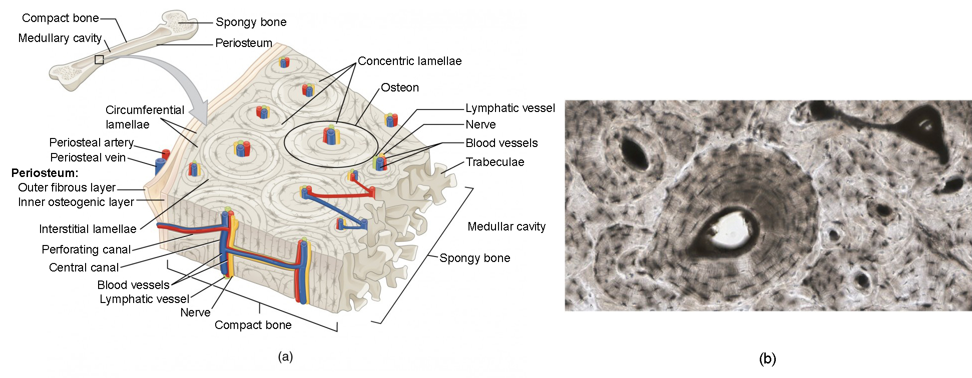
Bone Histology And Histopathology For Clinicians A Primer from slidetodoc.com Aorta , aldehyde fuchsin stain for elastin, 20x (extensive elastin in the wall). We have added a dotted line around the outside of the osteon in case you had trouble picking them out on the previous image. Note that the bone matrix is deposited in concentric layers called lamellae. In a cross section of a bone, you can usually see two types of bone tissues. Bone test anatomy and physiology 12 photos of the bone test anatomy and physiology anatomy and physiology bone lab test, anatomy and physiology bone markings test, anatomy and physiology bone practical test, anatomy and physiology bone tissue test, anatomy and physiology test on bone tissue, bone, anatomy and. Microscopic structure of compact bone consists of multiple cylindrical structural units known as osteons or haversian systems. Same bone section (slide 7) at higher magnification. The osteocytes are arranged in concentric rings of bone matrix called lamellae (little plates), and their processes run in interconnecting canaliculi.
Aorta , aldehyde fuchsin stain for elastin, 20x (extensive elastin in the wall).
The central haversian canal, and horizontal canals (perforating/ volkmann's) canals contain blood vessels and nerves from the periosteum. Bone cross section slide labeled : Cross section bone stock photos & cross section bone stock images. The tunic is secreted by the mantle, a thin cellular layer that also contains scattered bundles of muscle fibers and thus is capable of contraction. It can be found under the periosteum and in the diaphyses of long bones, where it provides support and protection. Clamp the section in a vise and carefully cut it to obtain a narrow slice. Obtain a demineralized compact bone preparation (in cross section), preferably from the diaphysis of a long bone, and prepare to examine it microscopically. 400x this image is from a different slide than the other two images on this page. Bone in arm pictures 12 photos of the bone in arm pictures bone cancer arm pictures, pictures of bone cancer in arm, bone, bone cancer arm pictures, pictures of bone cancer in arm The osteocytes are arranged in concentric rings of bone matrix called lamellae (little plates), and their processes run in interconnecting canaliculi. Cut the section to dimensions of about 5mm by 5mm chip. Histology spinal cord and ganglion : Very inneficient way to merge verticles.
400x this image is from a different slide than the other two images on this page. Bone, dried ground preparation, human. The basic unit of structure in compact bone is the osteon. Mammal compact bone slide, ground c.s. Aorta , aldehyde fuchsin stain for elastin, 20x (extensive elastin in the wall).
Bone Structure Anatomy And Physiology I from s3-us-west-2.amazonaws.com Cut the section to dimensions of about 5mm by 5mm chip. This slide contained a cross section of a very small bone, and you are looking at the entire thickness of the shaft of the bone. Each of these cylinders is called an osteon or. Clamp the section in a vise and carefully cut it to obtain a narrow slice. Histology spinal cord and ganglion : In the peripheral nervous system axons are bundled together in structures called nerves. A layer of connective tissue called the perineurium pn surrounds each fascicle. 400x this image is from a different slide than the other two images on this page.
The central haversian canal, and horizontal canals (perforating/ volkmann's) canals contain blood vessels and nerves from the periosteum.
Obtain a demineralized compact bone preparation (in cross section), preferably from the diaphysis of a long bone, and prepare to examine it microscopically. Clamp the section in a vise and carefully cut it to obtain a narrow slice. Each of these cylinders is called an osteon or. Related posts of cross section of human bone diagram bone in arm pictures. Try pressing the section on the slide to ensure that the layer of glue is as thin as possible. The tunic is secreted by the mantle, a thin cellular layer that also contains scattered bundles of muscle fibers and thus is capable of contraction. Consistency in fascicular organization of tibial nerve. To the left is muscle tissue, and to the right is bone marrow. Same bone section (slide 7) at higher magnification. This slide contained a cross section of a very small bone, and you are looking at the entire thickness of the shaft of the bone. Bone matrix and cells bone matrix osseous tissue is a connective tissue and like all connective tissues contains relatively few cells and large amounts of extracellular matrix. Very inneficient way to merge verticles. This is a cross section through.
The central haversian canal, and horizontal canals (perforating/ volkmann's) canals contain blood vessels and nerves from the periosteum. *none of the slide images above are shown at their actual scale. There are two slides to examine. The 40x image shows a cross section through four fascicles f that are part of a nerve. The basic unit of structure in compact bone is the osteon.
Description from www.dartmouth.edu Mammal compact bone slide, ground c.s. Bone, ground thin, human, longitudinal section digitalscope compare these two slides with each other and with webslide 74 identify in each slide: 400x this image is from a different slide than the other two images on this page. Consistency in fascicular organization of tibial nerve. Cross section bone stock photos & cross section bone stock images. Very inneficient way to merge verticles. Bone cross section slide labeled : The central haversian canal, and horizontal canals (perforating/ volkmann's) canals contain blood vessels and nerves from the periosteum.
In the peripheral nervous system axons are bundled together in structures called nerves.
Related posts of cross section of human bone diagram bone in arm pictures. Clamp the section in a vise and carefully cut it to obtain a narrow slice. In a cross section of a bone, you can usually see two types of bone tissues. The tunic is secreted by the mantle, a thin cellular layer that also contains scattered bundles of muscle fibers and thus is capable of contraction. In the center of each osteon is the central canal, a space that houses blood vessels and nerves that supply bone. In this image the bar indicates the location of decalcified compact bone. The basic unit of structure in compact bone is the osteon. Smartdraw includes 1000s of professional healthcare and anatomy chart templates that you can modify and make your own. This is a cross section through. The central haversian canal, and horizontal canals (perforating/ volkmann's) canals contain blood vessels and nerves from the periosteum. Notice the layered effect in the matrix. Microscopic structure of compact bone consists of multiple cylindrical structural units known as osteons or haversian systems. Cross section bone stock photos & cross section bone stock images.